-
Antibody Structure and Function
Antibodies, also known as immunoglobulins, are proteins shaped like a Y that the immune system makes to identify and counteract substances, like pathogens or harmful molecules. Their distinct design enables them to attach to their target antigen [1].
The fundamental structure of an antibody comprises two chains and two light chains connected by disulfide bonds. The variable sections at the ends of the Y shape are responsible for recognizing antigens while the constant parts carry out functions like opsonization, complement activation and neutralization. Antibodies possess a range of variability in their regions allowing them to recognize numerous antigens. This variability is achieved through rearrangements and somatic hypermutation during B cell development resulting in billions of antibody types.
In the response antibodies play a vital role, in helping remove pathogens and infected cells using various methods. They can block toxins, viruses and bacteria by binding to them and stopping them from entering host cells. Furthermore, antibodies can flag pathogens for destruction by cells through opsonization. Trigger the complement system—an array of proteins that can either directly destroy pathogens or call-in immune cells.
-
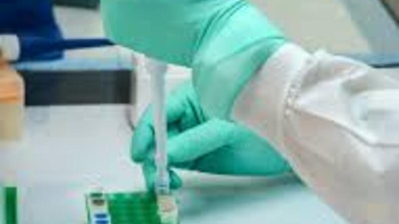
Immunoassay Techniques
Immunoassays are methods that make use of the bonding, between antibodies and target antigens for identifying and measuring specific target molecules. These techniques have brought advancements in fields like medical testing, environmental surveillance and scientific studies [2].
One of the most employed immunoassay approaches is the enzyme linked immunosorbent assay (ELISA). In this method antibodies are fixed on a surface and the interaction between the antigen and antibody is detected through an enzyme linked color reaction or fluorescence. ELISAs can be applied for both qualitative or quantitative assessment of molecules such as proteins, hormones, and infectious agents. These are also manufactured in the form of ELISA kits, where all the components that are required to perform the assay are provided. The advantage of purchasing a ready-made kit is that all the reagents have already been standardized to give accurate and reproducible results.
Another essential immunoassay technique is the Western blot, which separates proteins based on their size using gel electrophoresis before transferring them onto a membrane. Specific antibodies are then used to identify target proteins by their weight and antigen binding properties.
Immunohistochemistry (IHC) and immunocytochemistry (ICC) are methods that utilize antibodies to visualize proteins or antigens in tissue sections or cells. These techniques play a role in pathology as well as research endeavors, like investigating protein expression patterns and cellular locations.
Lateral flow tests, also known as tests (RDTs) are quick and convenient immunoassays that use antibodies fixed on a nitrocellulose membrane. These tests are commonly used to detect a range of substances, like agents, drugs and hormones making them valuable tools in healthcare facilities and field examinations.
-
Immunohistochemistry and Imaging Applications
Immunohistochemistry (IHC) and immunocytochemistry (ICC) are methods that use antibodies specificity to visualize and pinpoint tissue components. These techniques have become instruments in research, diagnostic pathology, and pharmaceutical development [3].
In IHC tissue samples are treated with antibodies that attach to target antigens such as proteins, enzymes or cell surface markers. These antibodies are then identified using fluorescent tags allowing for tracking and distribution of the target molecules within the tissue structure.
IHC plays a role in pathology by assisting in identifying tumor markers characterizing cell types and staging various diseases. For instance, IHC staining for estrogen receptor (ER) and progesterone receptor (PR) is regularly conducted in the diagnosis of breast cancer, for treatment planning purposes.
Apart, from using labeling, fluorescent immunohistochemistry (IF IHC) has become popular for its capability to detect targets simultaneously by utilizing different fluorophores. This method is especially valuable for examining the co localization and interactions among components.
Immunocytochemistry (ICC) follows a concept. Is applied to individual cells or cell cultures instead of tissue sections. ICC allows for the visualization and identification of proteins within compartments like the nucleus, cytoplasm or organelles.
-
Therapeutic Antibodies and Emerging Technologies
Therapeutic antibodies have emerged as treatment options for diseases beyond their diagnostic uses particularly in cancer treatment and autoimmune conditions [4].
Monoclonal antibodies (mAbs) are customized antibodies that target antigens with precision and affinity. These antibodies can be tailored to neutralize or inhibit the activity of disease-causing molecules, like growth factors or cytokines or to specifically attack and eliminate cancer cells that exhibit surface markers.
Several specialized antibodies have been given the light, for use transforming the way we treat different types of cancers like breast cancer, colorectal cancer and non-Hodgkins lymphoma. For instance, rituximab aims at CD20 in B cell lymphomas, trastuzumab targets HER2 in breast cancer and ipilimumab focuses on CTLA 4 in melanoma.
Antibody drug conjugates (ADCs) represent a group of treatments that merge the precision of monoclonal antibodies with the strength of drugs. In these combinations a potent drug is attached to an antibody that homes in on tumor markers. This method delivers the medication directly to cancer cells while reducing harm to tissues.
Bispecific antibodies (BsAbs) are specially designed to latch onto two markers opening up new avenues for treatment options. For example, BsAbs can bind to both a tumor marker and an immune cell receptor guiding cells to attack cancer cells (like blinatumomab for acute lymphoblastic leukemia).
Cutting edge technologies such as antibody engineering and display techniques (such, as phage display and yeast display) have broadened the range of antibody-based treatments available. These methods enable the creation of tailored antibodies, including those targeting complex markers or showing improved effectiveness.
Summary
Antibodies have completely transformed the field of research, diagnostics and treatment with their precision and adaptability. Techniques, like immunoassays and immunohistochemistry play a role in identifying various substances, disease indicators and cell components. The advancements in monoclonal antibodies antibody drug combinations and bispecific antibodies have notably enhanced the effectiveness of cancer treatments tailored to targets [5].
With progress in antibody engineering and presentation methods we can look forward to innovative applications based on antibodies. The future holds promising developments in diagnostics, treatments and scientific tools that utilize these biomolecules. Antibodies continue to drive the boundaries of science by providing valuable insights into disease mechanisms and leading to better health outcomes, for individuals.
References
[1] Schroeder, H. W., & Cavacini, L. (2010). Structure and function of immunoglobulins. Journal of Allergy and Clinical Immunology, 125(2), S41-S52.
[2] Wild, D. (Ed.). (2005). The immunoassay handbook. Nature Publishing Group.
[3] Ramos-Vara, J. A. (2005). Technical aspects of immunohistochemistry. Veterinary Pathology, 42(4), 405-426.
[4] Weiner, L. M., Surana, R., & Wang, S. (2020). Monoclonal antibodies: versatile platforms for cancer immunotherapy. Nature Reviews Immunology, 20(5), 317-327.
[5] Weiner, L. M., Surana, R., & Wang, S. (2020). Monoclonal antibodies: versatile platforms for cancer immunotherapy. Nature Reviews Immunology, 20(5), 317-327.